3D cone beam CT scanner in Boise, ID
Our in-office 3D cone beam CT scanner provides detailed three-dimensional imaging of your teeth, jaw, and surrounding structures in a single 20-second scan. More detail than traditional X-rays with less radiation.
Why 3D imaging matters for your dental care
3D visualization
Traditional dental X-rays produce a flat, two-dimensional image where structures overlap and details can be obscured. Our cone beam CT scanner captures a full three-dimensional volume of your teeth, jawbone, nerves, sinuses, and airway, giving your dentist a complete picture from every angle before planning treatment.
Lower radiation than hospital CT
A dental cone beam CT scan delivers significantly less radiation than a medical CT scan, typically 50 to 80 percent less. The focused beam targets only the area of interest, minimizing exposure to surrounding tissues. A single scan exposes you to roughly the same amount of radiation as a short domestic flight.
20-second scan
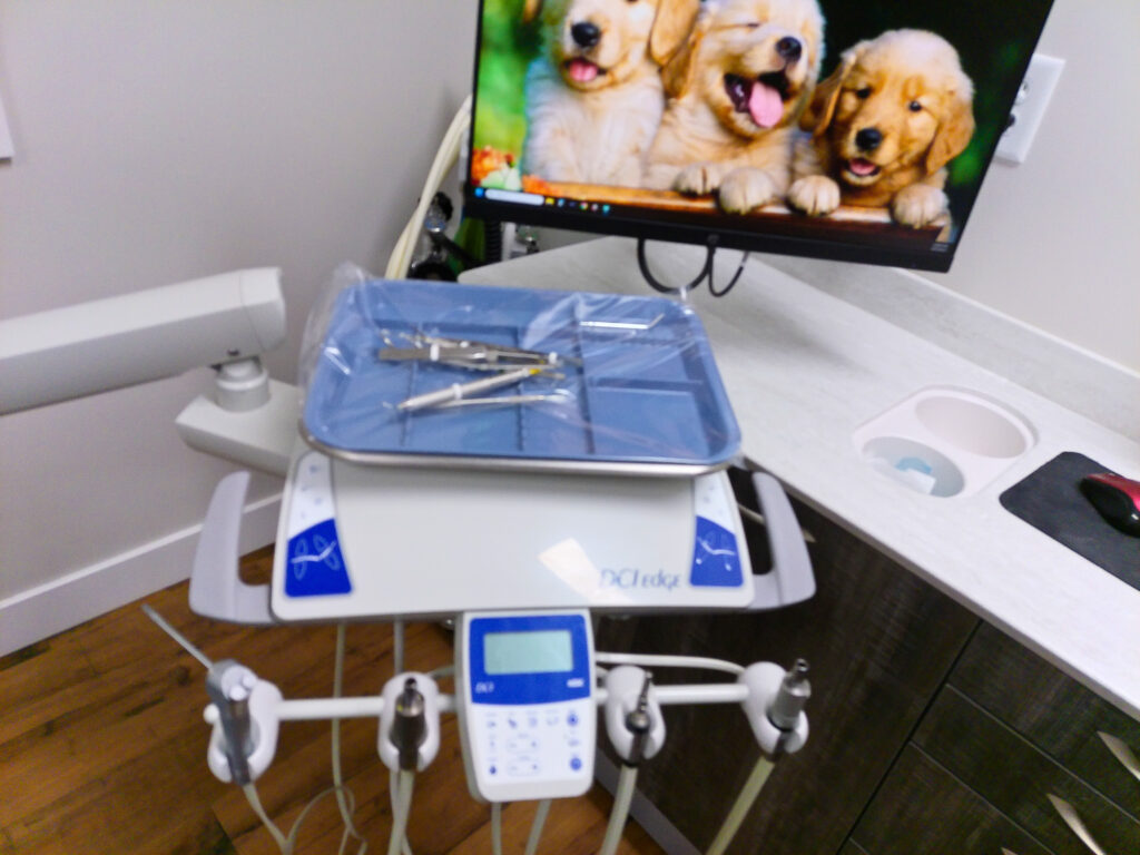
The entire scan takes approximately 20 seconds. You stand or sit comfortably while the scanner rotates around your head in a single pass. There is no enclosed tube, no loud noise, and no claustrophobia. The 3D image is available for review on-screen within minutes.
In-office, no referral needed
Our cone beam CT scanner is located right in our Boise dental office. You do not need a referral to an imaging center, a separate appointment, or a trip to the hospital. We capture the scan during your regular visit and review the results with you the same day, keeping your diagnosis and treatment plan on track without delays.
When we use 3D imaging
Not every dental visit requires a 3D scan. We use cone beam CT imaging selectively, when the additional diagnostic information will meaningfully improve your treatment plan and outcome. Common situations include:
- Dental implant planning, 3D imaging lets us measure bone density, width, and height at the implant site with sub-millimeter accuracy. We can visualize the proximity of nerves and sinuses and plan the exact position, angle, and depth of each implant before surgery begins. This precision is what produces predictable, successful outcomes.
- Wisdom teeth evaluation, impacted wisdom teeth can sit dangerously close to the inferior alveolar nerve that provides sensation to your lower lip and chin. A 3D scan shows the exact relationship between the tooth roots and the nerve canal, allowing us to plan the safest extraction approach and counsel you on risk.
- Root canal diagnosis, complex root anatomy, extra canals, and curved roots are difficult to see on standard X-rays. Cone beam CT reveals the full three-dimensional canal system so the treating dentist can locate and treat every canal, reducing the chance of missed anatomy and reinfection.
- TMJ assessment, for patients with jaw pain, clicking, or limited opening, 3D imaging provides detailed views of the temporomandibular joint. We can evaluate the condyle shape, joint space, and surrounding bone to identify degenerative changes, displaced discs, or other structural issues contributing to your symptoms.
- Airway analysis, the cone beam scan captures a cross-section of your upper airway, allowing us to evaluate for narrowing that may contribute to snoring or obstructive sleep apnea. This information can guide referrals to sleep medicine specialists and help determine whether oral appliance therapy might be beneficial.
How it works
Positioning
You stand or sit comfortably in front of the cone beam scanner. We position your head using adjustable guides so the area of interest is centered in the scan field. You bite gently on a positioning stick to stabilize your jaw. There is nothing to wear, nothing to swallow, and no preparation required. The entire setup takes about one minute.
20-second scan
The scanner arm rotates smoothly around your head in a single pass, capturing hundreds of individual images from every angle. The scan is completely painless, you simply hold still for approximately 20 seconds. There is no enclosed tube, no loud banging, and no contrast dye. Patients who are uncomfortable with MRI machines find cone beam CT perfectly tolerable.
Instant 3D rendering
The scanner software assembles the individual images into a detailed three-dimensional model of your teeth, bone, nerves, sinuses, and soft tissue within minutes. Your dentist can rotate, slice, and zoom into the 3D model from any angle, revealing anatomy that would be impossible to see on a traditional two-dimensional X-ray.
Doctor review with you
Your dentist walks you through the 3D scan on a chairside monitor, pointing out the specific findings that affect your treatment plan. You see exactly what we see, the bone structure, nerve locations, problem areas, and the rationale behind our recommendations. This visual review helps you understand your diagnosis and make informed decisions about your care.
Frequently asked questions
Is a cone beam CT scan safe?
Yes. A dental cone beam CT scan uses significantly less radiation than a medical CT scan, typically comparable to a set of four to eight standard dental X-rays, or roughly equivalent to one to two days of natural background radiation. We follow the ALARA principle (As Low As Reasonably Achievable), meaning we only recommend a 3D scan when the diagnostic benefit clearly outweighs the minimal radiation exposure. The scan is not recommended for pregnant patients as a precaution, consistent with standard guidelines for all dental radiography.
How is a cone beam CT different from a regular dental X-ray?
A standard dental X-ray produces a flat, two-dimensional image, like a shadow projected onto a wall. Structures overlap, and depth information is lost. A cone beam CT scan creates a full three-dimensional model that your dentist can rotate, slice, and zoom into from any angle. This means we can measure bone thickness precisely, see the exact path of a nerve, identify a hidden fracture, or map every canal inside a tooth root. For routine checkups, traditional X-rays provide all the information we need. For complex procedures like implant placement, wisdom tooth extraction, or root canal therapy, the 3D scan provides critical details that flat X-rays simply cannot show.
Does insurance cover a cone beam CT scan?
Many dental insurance plans cover cone beam CT scans when they are medically necessary for diagnosis or treatment planning, particularly for dental implants, impacted wisdom teeth, and complex endodontic cases. Coverage varies by plan, and some classify 3D imaging under the diagnostic benefit while others place it under the major procedures benefit. We verify your specific coverage before the scan so you know your out-of-pocket cost in advance. For uninsured patients, the scan is included in many of our treatment packages, and we accept our in-house membership plan.
Do I need a 3D scan for my visit?
Not necessarily. For routine checkups, cleanings, fillings, and most standard dental procedures, traditional digital X-rays provide all the information we need. We recommend a cone beam CT scan only when the three-dimensional information will change or significantly improve your treatment plan, typically for implant placement, impacted wisdom teeth, complex root canals, TMJ evaluation, or suspected jaw pathology. Your dentist will discuss whether a 3D scan would benefit you based on your specific clinical situation, and the scan is never ordered unless there is a clear diagnostic reason.
Patient Resources for 3D Cone Beam CT Imaging
Cone beam CT (CBCT) is regulated three-dimensional dental imaging used for surgical planning, endodontic diagnosis, and orthodontic evaluation. The organizations below publish guidelines on its appropriate use and safety.
- American Academy of Oral and Maxillofacial Radiology (AAOMR), the specialty body for dental imaging, including position papers on CBCT use
- American Dental Association, Cone Beam CT, ADA’s evidence-based guide on when CBCT is medically indicated
- FDA, Dental Cone Beam Computed Tomography, federal radiation safety guidance for CBCT imaging
- American Association of Oral and Maxillofacial Surgeons, surgical specialty guidance on CBCT in implant and oral surgery planning
- American Association of Endodontists, CBCT in Endodontics, specialty guidance on small-volume CBCT for root canal diagnosis
Advanced imaging for more precise dental care.
Insurance and Payment for 3D Cone Beam CT Scans
3D cone beam CT scans are classified as diagnostic imaging, often covered at 80 to 100 percent by insurance. Some plans limit diagnostic imaging to once every 3 to 5 years.
We accept Delta Dental, Blue Cross of Idaho, Cigna, Aetna, and 10+ other carriers. No insurance? Our in-house savings plan saves you 20–40% on every procedure. Learn about all financing and payment options.